
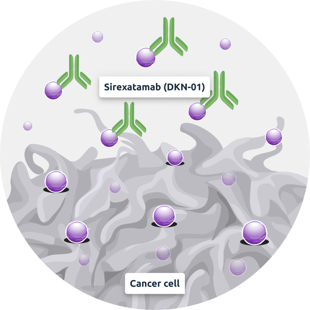
Sirexatamab (DKN-01)
– an anti-DKK1 antibody

Single agent and combination activity demonstrated in three different tumor types. Well tolerated as monotherapy and in combination with chemotherapy or checkpoint inhibitors.
Sirexatamab (DKN-01) binds and removes free DKK1 from the TME:

Reduces cell proliferation
Blocks signaling through CKAP4 and PI3K to downregulate Akt

Suppresses
MDSC cells
B-catenin dependent Wnt signaling reprograms MDSCs and reduces immunosuppressive activity

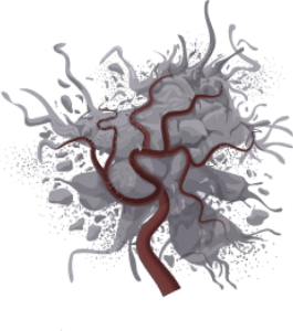
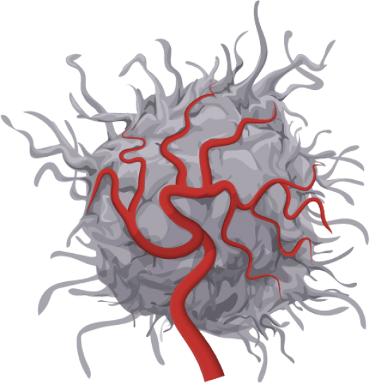
Reduces angiogenesis
Reduces blood vessel formation, upregulates key cytokines, IFNy, IL-15 and IL-33
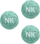
Activates
NK cells
Upregulates NK cell ligands on tumor, production of Granzyme B by activated NK cells

Sirexatamab (DKN-01) + anti-PD1
Sirexatamab (DKN-01) stimulates
innate immune system:

Stimulates activity
of NK cells

Induces
pro-inflammatory
tumor microenvironment

Upregulates PD-L1 (the target for PD-1 antibodies)
Anti-PD1 stimulate
CD8 T cell adaptive immunity:

Enhances tumor microenvironment for immune cells
to target and clear
the tumor
Activates
CD8 T cells
DKK1 expression determined using RNAscope

Chromogenic in situ
hybridization RNAscope
The biopsy sample is stained to identify DKK1 mRNA Pathologist determines histology score (H-Score), measuring DKK1 expression rather than protein itself.

Each red dot is an individual mRNA for DKK1.
Amount of cells and intensity of staining is converted to H-Score.
